Anatomy and Pathology
Clinical symptoms
Diagnosis
Treatment
Surgical Stabilisation
Atlantoaxial instability is excessive movement between the first two cervical vertebrae, the atlas (C1) and axis (C2) as a result of either bony or ligamentous abnormality.
Anatomy and Pathology
The first cervical vertebra or atlas (C1) is a bony ring consisting of anterior and posterior arch, connected by the two lateral masses that articulate with the second cervical vertebra or axis (C2). A vertically projecting odontoid process (Dens) from the Axis serves as a pivot for C1 to rotate. A transverse ligament fixed to the sides of the atlas secures the dens with close proximity to the anterior arch of the atlas. It provides stability in neck flexion and prevents anterior translation of the atlas on the axis. Other ligaments namely the alar, apical and accessory ligaments provide the secondary restraints of C1/2 articulation.
C1/2 instability may result from congenital malformation or syndromes, trauma, inflammatory arthritis, infection, malignancy deposits, osteoarthritic and some metabolic diseases.
Atlantoaxial rotatory subluxation is an isolated entity. It may occur with or without an initiating trauma. Non-traumatic etiologies include juvenile rheumatoid arthritis, infection of upper respiratory tract (Grisel Syndrome), and after surgical interventions of oral pharynx such as tonsillectomy. Conservative treatment is usually advised, as most of them will resolve spontaneously.
Os odontoidium is a rare anomaly. The etiology is controversial, whether it is congenital or developmental. An oval or round ossicle, smooth and sclerotic of variable size, locates in the position as the normal odontoid tip.
Atlantoaxial instability may also result from erosive synovitis that weakens the transverse ligament, such as in rheumatoid arthritis.
The cervical spine may be affected in C1/C2 instability because of narrowing of the spinal canal in the event of anterior displacement of C1 and the skull above, or any space occupying mass, such as hypertrophic synovium in the spinal canal.
Clinical symptoms
There may be torticollis, neck or occipital pain, tenderness and stiffness of the neck.
If cervical spinal canal is compromised by inflammatory process or malalignment, patient may have weakness and clumsiness of the extremities. Very occasionally, symptoms of vertebro-basilar insufficiency such as dizziness and fainting spells may occur.
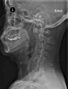
Diagnosis
On lateral X-ray of cervical spine, increased thickness of prevertebral soft tissue shadow may suggest local pathology at the atlantoaxial region.
The anterior atlantodens interval (AADI) should be 3.5 mm or less in adults. Dynamic instability can be documented by measurement in neutral, flexion and extension neck positions. If the space available for the cord behind the dens is less than 14 mm, neurological impairment may ensue.
CT scan may show more details about the bony pathology of the occipital-cervical junction. Sometimes, dynamic CT scan is taken to evaluate the C1/2 rotatory subluxation.
MRI is excellent for reviewing soft tissues and the status of neural elements. Apart from the bony compression, any soft tissue pathology like synovial mass can be assessed.
Treatment
Conservative treatment is usually advised for paediatric patients presented with atlantoaxial rotatory subluxation, and those with mild instability not accompanied with neurological symptoms or those rendered not fit for surgery.
Conservative methods comprise special caution in daily activities, avoidance of contact sport and serial clinical and radiological follow up to monitor the progress of symptoms. Sometimes, external immobilization means will be adopted if necessary, such as Halter traction, halo-body brace and different types of cervical orthosis.
Generally speaking, indications for surgery are those with acute or progressive neurological symptoms. Radiological monitoring of the atlantodens interval is useful to predict neurological deterioration.
Surgical Stabilisation
Atlantoaxial fusion with bone graft and posterior wiring had been used as early as 1939 to stabilise symptomatic C1/C2 instability.
Current advances in fixation include posterior C1-C2 clamp fixation, C1-C2 transarticular screws fixation or a combination of these procedures. Use of screws on the lateral masses or on the pedicels of C1 and C2 in combination with fixation rods are newer options.
The choice of procedures is much influenced by the reducibility of atlantoaxial subluxation, integrity of posterior ring of atlas and the expertise and preference of surgeons. However, rarely would occipital-cervical fusion be considered in congenital C1 /C2 instabiltiy, may be only in rare cases where the posterior arch of C1 is deficient.
Dr. WONG, Kam-kwong