Introduction
Musculoskeletal computed tomography (CT)
Equipment and Procedure
Advantages
Risks
Limitations
Introduction
Computed tomography (CT) is an imaging technique that allows cross-sectional images to be taken by using X-rays. It is a non-invasive, painless radiological test.
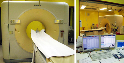
The patient lies on a scanning table, which slowly moves through a doughnut-shaped CT scanner. The scanner has an X-ray emitter on one side and a set of electronic X-ray detectors directly on the other side in a ring, called a gantry. Inside the gantry, the emitter emits X-rays and rotates around the patient in the axial plane at the level of interest. The electronic detectors will then receive the transmitted X-ray beam and calculate how much the beam is transmitted and how much the emitted X-ray is absorbed by the patient. The information is then analysed by a computer. Throughout the procedure, the scanning table moves the patient slowly in a constant speed and the process is repeated. Lastly, slices of cross-sectional images are then produced and displayed.
Due to a higher tissue contrast, CT images can provide a greater clarity of tissues within the human body than conventional images shown on X-ray films.
CT images are obtained using the same basic principles of plain radiography. As our body is made up of different types of tissues, there are different degrees of X-ray beam attenuation. High-density tissues like bone cause more attenuation (blocking) of X-ray beams and are therefore displayed as lighter (i.e. grey) than low-density tissues, such as muscles.
Musculoskeletal computed tomography (CT)
CT can provide important information when performed alone or in addition to other radiological investigations, such as plain radiography and magnetic resonance imaging (MRI). Depending on the situations, your doctor may consider ordering CT scans to help diagnosis and management.
Examples of clinical conditions suitable for CT assessment are
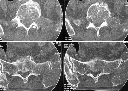
Managing trauma cases with fractures around a joint: acetabulum fracture (hip joint), tibial plateau fracture (knee joint), calcaneal fracture (subtalar joint in hind foot)
Managing trauma cases in areas difficult to be assessed with plain films alone, such as in the hand (carpal bone fractures) and in the foot (tarsal bone fractures)
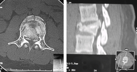
Spine
- detecting (or ruling out) spinal column damage
- assessing the extent of spinal column damage: burst fracture (thoracolumbar spine)
- assessment of spine: intervertebral disc
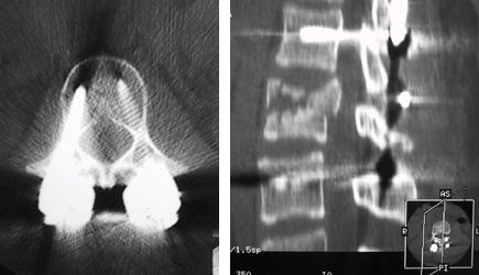
CTMM (myelogram): for patients contraindicated for MRI
Assisting the management of certain cancer cases
- assessment of osteoid osteoma
- diagnostic procedures such as biopsy of a deeply seated suspicious area
Assisting the management of certain cases with musculoskeletal infections
- detecting deeply seated abscess
- diagnostic/interventional procedure: aspiration of pus from abscess for diagnosis or placement of a drain tube for subsequent drainage
Identifying and locating radio-opaque foreign bodies
Identifying and locating radio-opaque loose bodies within a joint
Equipment and Procedure
Today, more technically advanced CT scanners are available. Helical (spiral) CT allows the X-ray emitter and detectors to rotate without stops as the patient passes through on the scanning table. Multidetector row CT (MDCT), also known as multislice CT (MSCT), has multiple detector rows instead of a single row of detectors as in a Helical CT scanner. This allows overlapping thin slices of data to be acquired in a much shorter period of time. The final CT images can be displayed in 3-D images as well as in sections of any desired plane. The shorter acquisition time opens up new areas for clinical use, especially in the field of cardiovascular-related medical conditions. It also greatly benefits certain patients, especially the elderly and the critically ill patients.
The patient must remain still on the scanning table while CT images are being scanned, as motion could result in blurred images. The computer workstation that processes the imaging information is located in a separate room. The whole procedure is painless. During the scanning process, as the emitter and detectors rotate within the gantry, you may hear slight buzzing and whirring sounds. You may experience discomfort from the cool temperature inside the CT room. With injury or pain, you may find lying on the hard scanning table uncomfortable. Your radiographer will assist you in finding the most comfortable position that still allows appropriate scanning. You will be alone in the exam room during the CT scan for a short period of time; however, the radiographer is able to see, hear and speak with patients during the whole scanning process through an audiovisual system and a monitor. The scanning procedure is usually finished within 10 minutes.
Advantages
CT imaging is a painless and non-invasive radiological investigation.
CT imaging can be performed even in patients implanted with medical devices.
With modern CT scanners, it is a rapid procedure and offers an accurate evaluation of bone and most soft tissues.
In emergency cases, CT imaging can quickly reveal internal injuries and bleeding.
CT imaging can provide real-time imaging, allowing minimally invasive interventional procedures to be performed with greater accuracy. These procedures include needle biopsies and aspirations.
Compared with plain radiography, CT images have a much better demonstration of anatomy with high contrast differentiation.
Compared with MRI, CT is more sensitive in detecting small amounts of tissue calcification and acute haemorrhage. CT scanners also generally have a lower installation and running cost when compared with MRI machines.
Risks
Computed tomography (CT) is an imaging technique that makes use of X-rays. Hazards are mainly related to irradiation.
Screening for pregnancy is essential. CT scanning is, in general, not recommended for pregnant women because of the potential risk to the fetus.
Contrast material may be used to further enhance image contrast. There is a risk of serious allergic reactions to contrast materials that contain iodine. Obtaining patients’ history of allergic reactions and other risk factors is essential if contrast material is going to be used during CT scanning.
Limitations
Compared with MRI, CT is less sensitive for obtaining excellent soft tissue image contrast. MRI can provide a better delineation of an injured spinal cord and the status of intervertebral discs.
Dr. YU, Kong-san