Anatomy
Cause
Symptoms
Types of fractures : 1. Fracture neck of femur
2. Intertrochanteric fracture
Preparing for surgery
Post surgical treatment
Complications
How the elderly can prevent falls
Anatomy
Osteoporosis is seen in most geriatric hip fracture cases. Other common osteoporotic or fragility fractures include:
- Fracture proximal humerus
- Fracture distal radius
- Fracture pelvis
- Collapse vertebrae
Geriatric hip fracture can usually be classified into two types:
Fracture neck of femur
Introchanteric fracture
Cause
Even slight injuries – like slipping from the bed or falling on the road, stumbling over the doorstep, slipping and hitting the hip while squatting down – can lead to fractures in the neck or trochanter of femur among the elderly. Other possible causes to elderly falls include: acute stroke, high blood pressure, poor eyesight, side effects of medication and the condition of the living environment.
Symptoms
Hip pain, difficulty in raising legs, in standing or in walking, as well as shortened and deformed limb turning outward are the common clinical symptoms of hip fractures.
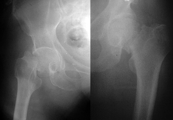
Types of fractures : 1. Fracture neck of femur
The fracture will damage the vessels that transmit blood to the neck of the femur, resulting in avascular necrosis of femoral head or nonunion. As such, femur head replacement will usually be employed for elderly patients. Reduction and internal fixation will apply to younger patients or cases when only mild dislocation occurs in the neck of the femur and the head part can be kept.
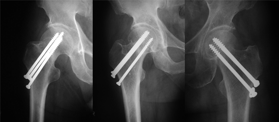
Surgical treatments
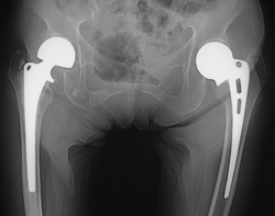
i. Hip screw Patients will receive surgery under general or local anesthesia. With the help of traction table and X-ray, doctors will perform reduction through a light incision made at the lateral thigh, using screws to lock the femur head firmly.
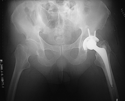
iii. Total joint replacement: performed under general or local anesthesia. Doctors will remove the damaged femur head and position a new metal one, whilst replacing the acetabulum with an artificial cup.
2. Intertrochanteric fracture
In most cases, the fracture will not damage the vessels that transmit blood to the femur head, and internal fixation will be the usual treatment option. There are stable and unstable intertrochanteric fractures, subject to the comminution level of the affected bone and different treatments will apply. Common surgical treatment options include:
Dynamic hip screw
Proximal femoral nail
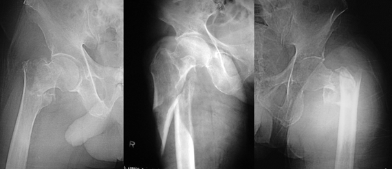
Surgical treatments
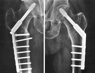
i. Dynamic hip screw Patients will receive surgery under general or local anesthesia. With the help of traction table and X-ray, doctors will perform reduction through a light incision made at the lateral thigh, using dynamic screws to lock the femur head firmly.
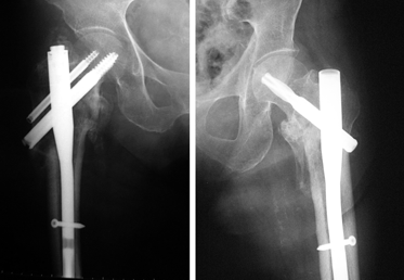
ii. Proximal femoral nail Patients will receive surgery under general or local anesthesia. With the help of traction table and X-ray, doctors will perform reduction through a light incision made at the lateral hip, using a femoral nail to lock the fractured bone firmly. Compared with dynamic hip screw, the use of a proximal femoral nail involves a smaller incision, a shorter operation time, less blood loss and an overall more stable performance.
Preparing for surgery
Sometimes skin traction will be applied to help ease pain
Patient’s blood is tested and matched and the patient is X-rayed
The patient’s existing illnesses are treated and stablised, e.g.: high blood pressure, diabetes, anemia and shortness of breath
The patient undergoes physiotherapy for muscular training and breathing exercises
The patient is allowed no food of eight hours before the surgery
The patient receives thorough skin cleansing and/or shaving of the surgical site
A Foley catheter may be necessary
Post surgical treatment
In general, patients can gradually start eating again after surgery
Anti-pain injection and pain relief medication will be used
If a Foley catheter is inserted during surgery, it may be removed after two to three days; a saline drip or blood transfusion may be required
Any drainage tube for the wounds can usually be removed in two to three days
After partial / total hip replacement surgery, doctor may use an abduction pillow for temporary fixation of lower limbs. It will be replaced by a sling later on, and patients can then move their limbs more freely
The patient will receive a course of physiotherapy exercises
When patients feel less pain, they can try sitting up. Patients who have undergone hip replacement will require a special hip chair to help sit properly and later they can start learning to walk again
Patients will be discharged from hospital in around two to three weeks
Complications
1. Common complications
Pressure sores caused by bed-bound
Venous thrombosis, pulmonary embolism
Infection and inflammation
Wound bleeding or hematoma
Improper wound healing
Recurrence of illnesses experienced before surgery, such as high blood pressure, stroke or diabetes
2. Complications specific to surgery
Complications related to hip screw / dynamic hip screw / proximal femoral nail surgery approach:
Bone screws / femoral nails loosen
Avascular necrosis of femoral head
Non-union
Malunion
Complications related to hip replacement approach:
Dislocation, subsidence and infection
Loosening
Sciatic nerve injury
Heterotopic ossification
How the elderly can prevent falls
Keep eye sight and hearing normal
For patients with high blood pressure, maintaining stable blood pressure to avoid dizziness is crucial
Use suitable walking support to treat leg injuries, knee pains or to keep balance: umbrellas and furniture are not reliable
Practice limb movements regularly or undergo balance training in a physiotherapy department
Alert and minimize household dangers, such as water, objects or small floor rugs; the passage to toilet should be clear of obstacles and well lit, especially at nighttime
People at a higher risk can consider wearing a hip protector
Prevent and treat osteoporosis (using both exercises and medication)
Dr. NGAI Wai-kit