Introduction
Symptoms
Diagnosis
Staging of disease
Treatment
Surgical Treatment
Surgical Treatment
Recent advances in surgical management of malignant bone tumours :
Introduction
The primary malignant musculoskeletal tumour is a relatively uncommon disease condition. In Hong Kong there are around 40 cases of primary malignant bone tumour and 130 cases of soft tissue sarcoma each year. Out of this, roughly 70% occur in the extremities and are managed in specialized tumour centers.
It should be noted, however, that the most common type of bone tumour is the metastases and these are malignancies that occur outside of the skeleton.
Symptoms
Many patients with malignant bone tumour will experience pain in the disease area, with or without swelling. The pain may or may not be related to activities and it can even wake the patient up at night. When pain is severe, it can possibly be due to a fracture in the tumour laden bone.
"Soft tissue sarcoma" on the other hand is a growing mass that is often painless. A rapid increase in size within a short period is an alarming sign.
Diagnosis
Patients with the above symptoms should seek a doctor’s advice. After a physical examination, an X-ray and looking at the patients history, the doctor should be able to decide whether there is a suspicion of malignancy.
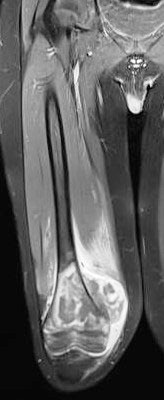
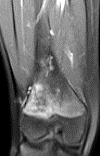
Further imaging such as MRI is an important step in diagnosis. When both clinical & imaging studies point to a suspicion of malignant tumour, it is essential to obtain histological diagnosis before surgical treatment. Doing surgery without adequate imaging and histological information is something to be discouraged. This is because such unplanned surgical excision often requires repeat surgery of a much bigger magnitude and even compromises the final outcome or survival of the patient.
Nowadays, a well established method to obtain histological diagnosis is image-guided needle biopsy that, in experienced hands, carries a diagnostic accuracy of over 95%.
Staging of disease
After establishing the diagnosis of malignant tumour, the next step is doing systemic staging studies. Traditionally this includes CT scans of the thorax as well as bone scans for detecting pulmonary and bone metastasis respectively. Positron emission tomography (PET) scans are also useful in certain situations but their role in the staging of primary malignant musculoskeletal tumours is not yet well-established.
Treatment
The treatment of primary malignant musculoskeletal tumour requires highly specialized multidisciplinary care by the orthopaedic surgeon, oncologist, paediatric oncologist, radiologist & pathologist. Generally speaking treatment consists of surgical excision with or without adjuvant therapy depending on the histological type and staging of the tumour.
Surgical Treatment
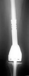
1. Limb-sparing surgery with tumour excision and reconstruction: This is possible when the tumour is low grade, or when it is diagnosed early enough for the anatomical extent of the tumour to allow excision with an adequate margin.
For soft tissue tumour, soft tissue defects may require tissue flap reconstruction. For bone tumours, bone defects may be reconstructed using bone grafting or metallic implant / mega-prosthesis.
2. Amputation is often needed in high grade tumours, where excision with an adequate margin is technically not possible, e.g. when the major neurovascular bundle is effected by the tumour.
Surgical Treatment
1. Limb-sparing surgery with tumour excision and reconstruction: This is possible when the tumour is low grade, or when it is diagnosed early enough for the anatomical extent of the tumour to allow excision with an adequate margin.
For soft tissue tumour, soft tissue defects may require tissue flap reconstruction. For bone tumours, bone defects may be reconstructed using bone grafting or metallic implant / mega-prosthesis.
2. Amputation is often needed in high grade tumours, where excision with an adequate margin is technically not possible, e.g. when the major neurovascular bundle is effected by the tumour.
Recent advances in surgical management of malignant bone tumours :
1. Computer assisted navigation
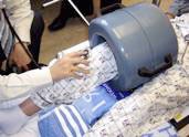
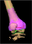
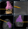
Sometimes it is important to be able to carry out very precise bone excision with the help of computer navigation. e.g. in joint-preserving excision, or in difficult anatomical areas like the pelvis or sacrum.
CT & MRI imaging data are used for pre-operative 3D planning of the surgery. During operation, real time navigation of surgical tools is then utilized for precise execution of the bone cuts according to pre-operative plan within a few millimeters of accuracy. (figure 1)
2. Growing prosthesis in very young children with malignant bone tumour
By including a growing mechanism into a prosthesis, the length of the affected limb can be extended gradually to allow equalization of leg length while the child is growing. The most sophisticated growing prosthesis currently available is the non-invasive grower where prosthesis lengthening is achieved by a magnetic coil without the need of additional surgery. (figure 2)
Dr. Timothy SO